Breast cancer is probably one of the most feared diagnoses a woman can get. The figures are undoubtedly scary, so what can we do to reduce our risks? Well, the good news is there are a lot of measures we can take.
Roula Allam talks to Dr Reine Nader, a Lebanese specialist in Diagnostic Imaging and Radiology with a diploma in Interventional Radiology and Cancerology from Versailles University, about effective prevention tactics. Plus, she asks Nader, who also has Specialised Medical Training (DFMS) diplomas from Antoine-Béclère Hospital, Gustave Roussy Institute of Oncology, Sorbonne University and Pierre and Marie Curie University, which women are more likely to be at risk.
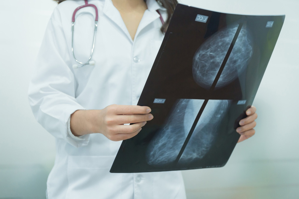
Are clinical breast exams as important as mammograms?
A clinical exam is an important step to detect breast anomalies. Most of the time the palpable lesions are benign, however, the percentage of breast cancer detected from a palpable lesion is much higher than the percentage spotted from a mammography. When faced with a palpable lesion, an evaluation should be done to exclude a malignant lesion. The clinician discovers a palpable lesion and addresses the patient for a diagnostic investigation, or it is discovered by self-examination, when the patient discovers the anomaly and goes to the physician. The radiologist may also discover a lesion while palpating the breast before the mammography. The clinical exam is 54 percent sensitive and 94 percent specific. It is effective and can detect up to 29 percent of cancers not seen by the mammogram. It is done in an illuminated room, with the patient in the orthostatic position, sitting with elevated arms. Before menopause, it is best done one week after your period. So yes, of course a clinical exam is as important as a mammogram and sometimes even better.
At what age should we begin having mammograms and how often?
In France, we begin to invite women for mammograms at the age of 50 if there aren’t any known risk factors, and it is done every two years till the age of 74. In Lebanon, we see most of the physicians asking patients to start having a mammography at the age of 40 and to repeat it every year. In my opinion, if the patient doesn’t have a risk factor, you can start having mammograms at the age of 50. Then depending on the findings, we can decide when it should be repeated.
Do self-exams really help?
As I previously said, a self-exam is a part of a clinical exam and it plays a major role in detecting lesions, especially if women do it regularly. Each woman knows her breasts, their form and shape and can detect a change and palpate any tiny lesion. I have seen lots of patients who came to me after a self-exam. Their lesions were found and treated early on and they are now cured. So a self-exam is definitely a major step in detecting breast lesions.
Are active women less likely to develop and die from breast cancer?
Physical activity is a protective factor against breast cancer, even if it starts later in life. Studies have shown it decreases the risk by 20 to 40 percent. Plus active women have a healthier lifestyle, which is also a defense against breast cancer. They are also more aware of any changes in their breasts and seek medical care and treatment early, so they are less prone to dying from breast cancer.
Is being overweight risky, especially after menopause?
Yes, in fact the intake of saturated fat before menopause increases the relative risk (RR) of breast cancer (RR, 1.25). In menopausal women, a fibre-rich regime protects from breast cancer (RR, 0.85). Obesity is a risk factor after menopause; it is related to the estradiol hormone level, which is elevated in these women, rather than being related to the obesity itself.
What are the most common signs of breast cancer?
Some of the most common signs include a palpable lesion, asymmetry of the breasts, unilateral ptosis and nipple or breast retraction. Other signals include discharge (fluid) from the nipple, inflammation or redness, skin changes and edema. Small anomalies are more evident when leaning down and elevating the arms.
How much does a family history really matter?
A family history is one of the biggest risk factors when it comes to breast cancer and it largely depends on how related you are. If you are a first-degree relative, the relative risk is 2.1: mother (RR, 2), sister (RR, 2.3) and daughter (RR, 2.1). If two first-degree relatives are diagnosed with breast cancer then the risk is higher (RR, 3.6). If a second-degree relative is diagnosed it is lower (RR, 1.5). Plus this is related to the transmission of Brca1 and Brca2 mutations in an autosomal dominant mode and many other genetic syndromes and mutations.
What other factors affect our risks?
Aspects like age, at what age you started puberty, when you begin menopause and parity, as well as the age of the first delivery are considered. Additionally, a woman’s body mass index, personal cancer history and a history of thoracic irradiation before the age of 30 can have an effect on your chances of having breast cancer. Hormonal treatment for menopause, your lifestyle, atypical lesions and breast density can too.
So why are women with dense breasts more likely to get breast cancer?
They are more likely to have breast cancer because first of all a dense breast (BI-RADS 3 or 4) is more likely to mask the lesions on the mammography. Due to the high density of the breasts, a lesion can be missed on the mammography. The second, and most important reason, is because they have more breast tissue than fat, so they are more likely to develop breast cancer. A mammography should be coupled with a breast ultrasound in this case.
What can we do if we are at high risk?
If you are in the high-risk category, you should start having a mammography at age of 30, even 25, coupled with an annual breast MRI. If there is a genetic mutation, an MRI should be done between the ages of 25 and 29, then you should alternate between an MRI and mammography every six months once you hit 30. Finally, there’s the option of a prophylactic mastectomy for the patients who are at high risk and don’t wish to go through the entire stressful experience of investigations and detection.